
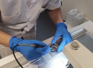
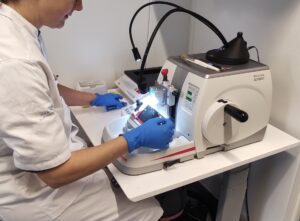
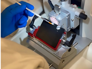
We process tissue/biopsies from patients in our own laboratory, as we have the equipment and expertise to fix, embed, and section tissue for further analysis. We perform tissue staining, including H&E staining, allowing our pathologist to evaluate the tissue and determine the tumor’s type, grade, and growth pattern.
The ability to conduct this research is crucial and plays a significant role in improving cancer treatment and survival rates.
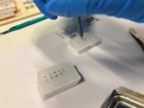
We perform immunohistochemistry using fluorescence biomarkers, a technique that visualizes and identifies specific cells in tissue sections by using antibodies labeled with fluorophores. We apply this technique in singleplex, duplex, and multiplex formats, helping us gain insights into cell localization, morphology, interactions between cells/proteins, and biological processes within the tissue.
This method provides us with a better understanding of an individual patient’s tumor composition and aggressiveness while also helping us identify the best treatment options. Ultimately, this can lead to more effective treatments with fewer side effects and improved prognoses.
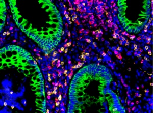
⬆ Colorectal adenoma: PanCK (epithel cells) GREEN – SYTO13 (kernels) BLUE – CD3 (T-cells) RED – CD8 (cytotoxic cells) YELLOW
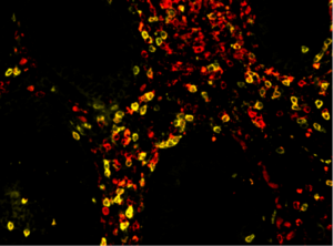
⬆ Colorectal adenoma: CD3 (T-cells) RED – CD8 (cytotoxic cells) YELLOW
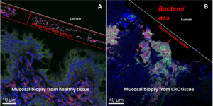
Fluorescence in situ Hybridization is a molecular biology technique where we use fluorescence-labeled probes that bind to RNA to visualize bacteria in tissue. We primarily examine a universal bacterial probe, as well as Bacteroides fragilis and Fusobacterium nucleatum. These bacteria can significantly impact cancer progression, and we investigate whether their presence, along with other bacteria, influences patient outcomes.
NanoString is a platform that utilizes advanced transcription and protein analysis to understand tumor-specific gene expression in tissue and blood.
We use specific immunohistochemistry panels on patient tissue, enabling us to map immune cell profiles and assess the tumor microenvironment. This allows for a better biological understanding of which patients may benefit from personalized treatments.
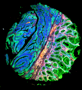
GeoMx technology can be used to analyze patient tissue stained with these immunohistochemistry panels. By utilizing fluorescence-labeled biomarkers, we generate an image of the tissue’s structure and select Regions of Interest within the tissue for digital RNA or protein quantification using specific probes. Subsequently, gene expression in different regions can be compared, and data analysis can be performed.
The same transcriptional analysis can be performed on isolated RNA from tissue or blood using NanoString kits with specific probe sets that bind to RNA sequences coding for particular genes. Gene expression analysis is conducted using the nCounter system, helping us understand how individual patients respond to specific interventions or treatments.
TriMeth is a blood-based DNA methylation analysis that enables the detection of colon cancer with high specificity and sensitivity using dPCR. This technology uses cfDNA from plasma to assess methylation levels in genes of interest, detected through specific primers and probes.
This method supports early diagnosis and contributes to predictive models for immunotherapy responses in individual patients. In a research context, it is a valuable platform that aids in the development of personalized medicine, ultimately benefiting patients.
Patient-derived organoids (PDOs) are three-dimensional cell cultures that we grow from patient tissue samples, including biopsies from both healthy organs and tumors, such as colorectal and pancreatic cancer. The tissue is dissociated into single cells and embedded in a gel, which, along with specialized medium, plays a crucial role in enabling the cells to form their three-dimensional structure.
Compared to traditional cell lines, PDOs provide a more physiologically relevant model of the organ, preserving the genetic and phenotypic complexity of the original tissue. This makes them an ideal model for personalized medicine, with promising applications in cancer research, including drug response testing and tailored treatments.
Like conventional organoid culture, Air-Liquid Interface (ALI) models utilize patient-derived tissue. In this model, small tissue fragments are cultured in a dual-phase environment, where the upper side is exposed to air, and the lower side to liquid. This setup closely mimics the physiological conditions of the intestinal epithelium, making it particularly suitable for colorectal cancer research.
A key advantage of this model is that the patient’s native microenvironment, including immune cells, is preserved in the culture. This enables us to test personalized immune responses, which can be a crucial tool in cancer research.
Phagocytosis assays are an experimental method used to characterize the ability of phagocytes to engulf and degrade, among other things, bacteria and apoptotic cells. In our lab, we perform this method on immune cells that are isolated from the patient’s PBMCs derived from a blood sample. We use this method as a tool to assess the patient’s immune response to a given treatment. To analyze the cells’ ability to phagocytose, we use both confocal microscopy and fluorescence measurement.
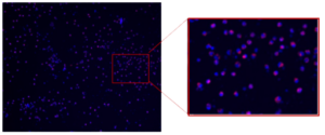
⬆ Phagocytosis assay on monocytes isolated from PBMCs. DNA is stained with Hoechst and visualized in blue. Phagocytosed E. coli bacteria are labeled with a red fluorophore and therefore visualized in red.